
© Dennis Kunkel Microscopy
Home Resources Pathogens What are Acanthamoeba spp. and Naegleria fowleri?
What are Acanthamoeba spp. and Naegleria fowleri?
© Dennis Kunkel Microscopy
These organisms are protozoa commonly referred to as pathogenic free-living amoebae. They are ubiquitous in aquatic and soil habitats.
What diseases are caused by Acanthamoeba spp. and Naegleria fowleri?
Several species of Acanthamoeba can invade the brain and meninges of immunocompromised individuals and cause granulomatous amoebic encephalitis. Acanthamoeba usually enters the body through a cut, wound, or through the nostrils. The amoebas then spread via the bloodstream to other parts of the body such as the lungs, brain, and spinal cord. Symptoms include headaches, stiff neck, nausea and vomiting, tiredness, confusion, lack of attention to people and surroundings, loss of balance and bodily control, seizures, and hallucinations. The mortality rate associated with Acanthamoeba infection is very high.
Naegleria fowleri causes the disease primary amebic meningoencephalitis (PAM). Infection occurs by the organism entering the body through the nose then traveling to the brain and spinal cord. This generally occurs when people use warm freshwater for activities such as swimming or diving. The initial symptoms include headache, fever, nausea, vomiting, and stiff neck. Later symptoms include confusion, lack of attention to people and surroundings, loss of balance, seizures, and hallucinations. The disease progresses rapidly and usually causes death within 3 to 7 days.
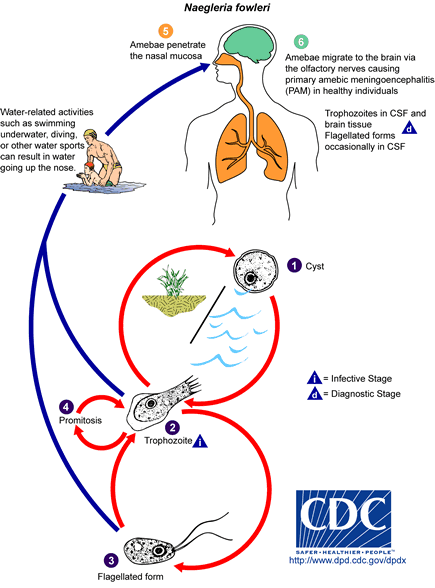 Naegleria fowleri has three stages, cysts (1), trophozoites (2), and flagellated forms (3) , in its life cycle. The trophozoites replicate by promitosis (nuclear membrane remains intact) (4).
N. fowleri is found in fresh water, soil, thermal discharges of power plants, heated swimming pools, hydrotherapy and medicinal pools, aquariums, and sewage. Trophozoites can turn into temporary non-feeding flagellated forms which usually revert back to the trophozoite stage.
Trophozoites infect humans or animals by penetrating the nasal mucosa (5) and migrating to the brain (6) via the olfactory nerves causing primary amebic meningoencephalitis (PAM).
N. fowleri trophozoites are found in cerebrospinal fluid (CSF) and tissue, while flagellated forms are occasionally found in CSF. Cysts are not seen in brain tissue.
Who is more susceptible to Acanthamoeba spp. and Naegleria fowleri?
People with compromised immune systems or those who are chronically ill are at greatest risk for Acanthamoeba infection. Healthy individuals have been shown to develop Naegleria fowleri infection. The highest risk of Naegleria fowleri infection is during the summer months and among those who participate in recreational freshwater activities.
Epidemiology of Acanthamoeba spp. and Naegleria fowleri
Both of these organisms are distributed globally in the environment, however, they rarely cause disease. To date there have been over 100 cases of granulomatous amoebic encephalitis in immunocompromised individuals caused by acanthamoeba infection. More than 160 cases of primary amoebic encephalitis have been recognized in healthy individuals. These infections have occurred in many countries on all inhabited continents.
Incubation Period
The incubation period for acanthamoeba infection is unknown but is probably from weeks to months. The incubation period for Naegleria fowleri infection is 3 to 7 days.
Diagnosis of granulomatous amoebic encephalitis and PAM
Suspected cases of granulomatous and primary amoebic encephalitis are diagnosed by microscopic examination of fresh cerebrospinal fluid for motile amoeba. Stained smears of cerebrospinal fluid can also be examined microscopically.
Treatment of granulomatous amoebic encephalitis and PAM
The primary treatment for Naegleria fowleri infection is the antifungal medication amphotericin B. It is usually injected intravenously or into the space around your spinal cord. However, most infections are difficult to treat successfully. Several drugs have shown effectiveness against granulomatous amoebic encephalitis. Unfortunately treatment is not standardized and is limited.
Contact EHA Consulting Group today for more information about how we can assist your company.